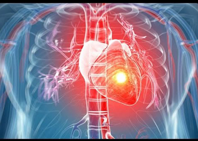
***Device Closure procedures in Navi Mumbai*** provide specialized treatment for congenital heart defects including ASD, VSD, and PDA. Our ***expert cardiac device closure services in Navi Mumbai*** utilize advanced minimally invasive techniques to correct these birth defects, restoring normal heart function and improving quality of life for patients.
Atrial Septal Defect (ASD) Treatment in Navi Mumbai: This is a congenital heart defect in which blood flows between the atria (upper chambers) of the heart. A wall called the Interatrial Septum separates the Atria. If this septum is defective or absent, then oxygen-rich blood can flow directly from the left side of the heart to mix with the oxygen-poor blood in the right side of the heart, or vice versa. Our ASD closure specialists in Navi Mumbai address this condition to restore normal oxygen levels in the arterial blood that supplies the brain, organs, and tissues.
Ventricular Septal Defect (VSD) Treatment in Navi Mumbai: This is a defect in the ventricular septum, the wall dividing the left and right ventricles (lower chambers) of the heart. The extent of the opening may vary from pin size to complete absence of the ventricular septum, creating one common ventricle. Our VSD closure procedures in Navi Mumbai address defects in the ventricular septum, which consists of an inferior muscular and superior membranous portion and is extensively innervated with conducting cardiomyocytes (cardiac muscle cells).
Patent Ductus Arteriosus (PDA) Treatment in Navi Mumbai: This is a condition wherein the ductus arteriosus (fetal blood vessel) fails to close after birth. This vessel does not close and remains "patent" (open), resulting in irregular transmission of blood between the aorta and the pulmonary artery. Our PDA closure services in Navi Mumbai are particularly important for newborns with persistent respiratory problems such as hypoxia and have proven essential for premature newborns due to underdevelopment of the heart and lungs.